Coral Infectious Diseases: Brown Jelly Disease and Other Soft Tissue Problems and Management
An overview of infectious tissue problems seen in LPS corals and other corals with substantial soft tissue, including brown jelly disease, with considerations for early detection and prevention of spread.
Key Takeaways
An overview of infectious tissue problems seen in LPS corals and other corals with substantial soft tissue, including brown jelly disease, with considerations for early detection and prevention of spread.
What is Brown Jelly Disease
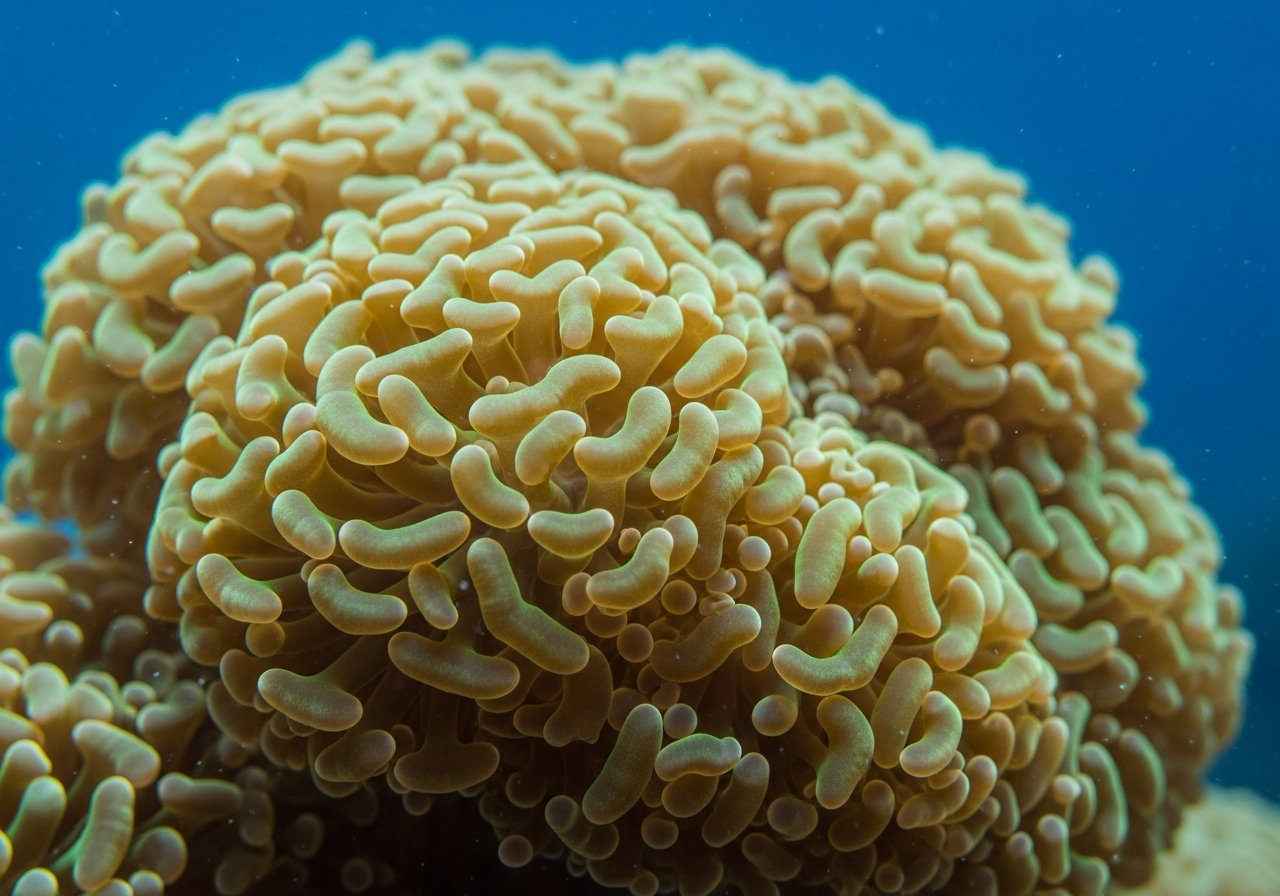
Brown Jelly Disease refers to a condition in which coral tissue dissolves into a brownish jelly-like substance. It is particularly common in LPS corals with thick soft tissue, such as Euphyllia species exemplified by hammer corals, torch corals, and frogspawn corals. The disease is thought to develop when bacteria or protozoa opportunistically invade and proliferate in tissue that has been damaged or weakened for some reason. While it is rarely seen in healthy tissue, once it takes hold, the infection can spread to surrounding tissue at a remarkably rapid rate—sometimes within hours to a day. This is a soft tissue-specific problem that should be distinguished from RTN and STN (Rapid and Slow Tissue Necrosis), which affect the skeletal tissue of SPS corals and differ in both appearance and progression mechanisms (see RTN・STN in SPS Corals: Causes and Management). Similar-looking symptoms are reported not only in hammer and torch corals but also in other LPS corals with thick soft tissue and some soft corals, where tissue dissolves through the same mechanism. It is best not to narrowly characterize this as "a disease unique to Euphyllia."
Common Triggers for Onset
Physical damage from transport or fragging, stress from sudden changes in water quality or temperature, and tissue weakening from prolonged poor water conditions are common triggers for Brown Jelly Disease. Additionally, tissue damage from predation by nudibranchs, planarians, and other aquarium pests can create entry points for infection (see Coral Pest Control Guide). Newly introduced corals that have not yet acclimated to their environment, or corals that have experienced prolonged transport stress, warrant particularly close observation during this critical period.
Key Signs for Early Detection
If you notice part of the tissue appearing brown and gelatinous, observe an unusual slime coating or membrane formation, or see tissue rapidly receding, these are warning signs of Brown Jelly Disease. Unlike bleaching (where tissue color fades but the tissue itself remains; see Coral Bleaching: Causes and Recovery), the defining characteristic of this disease is the rapid loss of tissue itself. Maintaining a keen eye for your corals' normal appearance through daily observation makes it easier to spot such changes early.
Initial Response to Prevent Infection Spread
When a affected coral is suspected, common practice is to physically separate it from other corals or, if possible, move it to a quarantine container. There is concern that the gelatinous tissue or water that has contacted it could spread infection to other healthy corals. Some aquarists carefully remove only the visibly affected portions using clean tools and preserve the healthy tissue, but this judgment depends on the severity and type of symptoms and cannot be universally recommended. If you are uncertain about the decision, or if the symptoms are widespread, it is prudent to avoid self-treatment and instead consult an aquarium specialist or an experienced aquarist early on.
Preventive Measures
Establishing a quarantine routine—isolating newly acquired corals from others and observing them for a period of time before introducing them to the main tank—is fundamental in preventing infectious problems. Quarantine containers should use separate water and equipment from the main tank; use this time to check for tissue condition and signs of predation damage before moving the coral to the main aquarium. When fragging or relocating corals, handle them gently to minimize tissue damage, and maintain stable water quality and temperature to reduce chronic stress. Regarding coral spacing, refer to LPS Coral Placement and Coexistence and arrange corals to avoid contact and predation-related injuries. Because early signs of Brown Jelly Disease can occasionally resemble tissue damage from pest predation, regularly checking for the presence of pests allows for easier identification of the underlying cause.
Keeping Treatment Expectations Realistic
While early detection and response to Brown Jelly Disease and other infectious problems can increase the chances of minimizing damage, there is no guarantee of complete recovery or prevention of spread. Depending on the speed of progression and the condition of the coral, treatment may not arrest the disease in time, and the coral may lose much of its tissue. While excessive pessimism is not warranted, a more pragmatic approach is to base your strategy on daily observation habits and early consultation with specialists, rather than searching for a "guaranteed cure." Cultivate a willingness to consult aquarium specialists and veterinarians who can treat marine organisms.